

Before cataract surgery, I’d ask one question: Are my eye’s surface and lens measurements stable? Dryness can affect lens calculations – even when my eyes feel fine. Treatment may take weeks to months, so feeling better isn’t the only sign that I’m ready.
I’d use these 5 steps to prepare:
- Report symptoms and contact lens use. Have my cornea, tear film, and eyelids checked.
- Check measurement accuracy. Ask whether tear stability and corneal shape could affect my lens choice.
- Follow my treatment plan. Use prescribed drops and eyelid care, and address infection, inflammation, or eyelid problems.
- Return for repeat testing. <u>Wait for reliable readings</u> before finalizing lens calculations.
- Plan care before and after surgery. Confirm medications, costs, follow-up visits, and whom to call with concerns.
My surgeon decides when it’s safe to proceed. Sudden vision loss or severe eye pain needs urgent eye care.
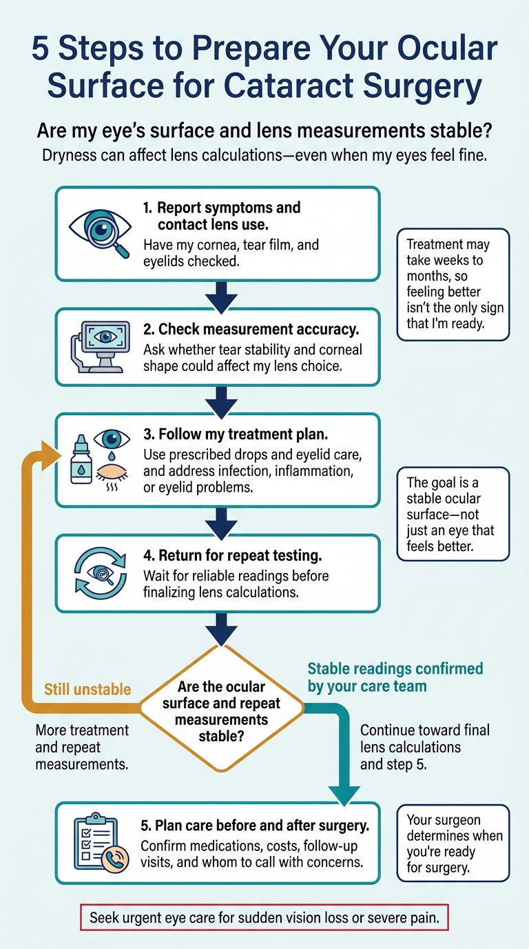
5 Steps to Prepare Your Ocular Surface for Cataract Surgery
Setting the Stage for Success: Evaluating & Preparing the Ocular Surface Before Cataract Surgery
sbb-itb-5ef6ec9
Checklist 1: Report Symptoms and Get an Ocular Surface Exam
Before taking final cataract measurements, review symptoms and contact lens use, then examine the eye’s surface.
Share Symptoms and Contact Lens Use
Patient checklist:
- [ ] Symptoms: Report any eye discomfort, even if it’s mild.
- [ ] Contact lenses: Share whether you currently wear contact lenses and whether they cause discomfort.
Check the Cornea, Tear Film, and Lid Margins
Clinician checklist:
- [ ] Corneal surface: Check for staining, epithelial defects, irregular epithelium, and corneal filaments.
- [ ] Tear film: Assess tear stability and how well tears cover the surface between blinks.
- [ ] Lid margins and meibomian glands: Check for blepharitis, meibomian gland dysfunction (MGD), and signs of Demodex mites.
- [ ] Active infection or inflammation: Look for blepharitis, hordeola, or significant inflammation.
- [ ] Lid structure: Check for misdirected lashes, abnormal lid position, and scarring.
Decision Table: Proceed, Treat, or Reassess
| Finding Category | Clinical Observation | Action |
|---|---|---|
| Normal Surface | Clear cornea, stable tear film, healthy lids | Proceed with measurements and surgery |
| Mild Disease | Minor staining or dryness that does not affect vision | Start treatment; may proceed with caution |
| Unstable Surface | Irregular epithelium, unstable tears, significant staining | Treat first; repeat measurements once the surface is stable |
| Active Infection | Blepharitis, hordeola, or significant inflammation | Delay final planning until infection or inflammation is controlled |
| Structural Issues | Abnormal lid position, misdirected lashes, scarring | Address structural problems before final planning |
If findings are inconsistent, confirm tear stability before choosing final IOL measurements.
Checklist 2: Check Tear Stability and Measurement Accuracy
After the surface exam, your team checks whether your measurements are stable enough to plan your lens selection.
Your clinician selects tests based on your exam findings and lens goals. Tell your surgeon if you want a toric or multifocal lens. Both options depend on accurate measurements.
Test Table: What Each Test Shows
| Test | Finding evaluated | Use in surgical planning |
|---|---|---|
| Corneal Topography | Corneal shape and regularity | Identifies irregular corneal shape that may affect lens choice. |
| Manual/Automated Keratometry | Corneal curvature and astigmatism | Guides IOL power and toric lens calculations. |
| Optical Biometry (e.g., IOL Master) | Eye length | Guides IOL power calculations. |
| Verion System | Astigmatism, pupil center, and visual axis | Guides astigmatism correction and lens alignment. |
| ORA System | Intraoperative refractive guidance | Helps guide the final lens selection during surgery. |
If results are inconsistent, your team treats the ocular surface before taking measurements again.
Checklist 3: Follow Your Prescribed Treatment Plan
If your eye’s surface is unstable, treatment comes first. The goal is a stable ocular surface – not just an eye that feels better. Follow your prescribed plan even after symptoms improve.
Support the Tear Film and Treat Eyelid Disease
- [ ] Use prescribed lubricating drops, lid hygiene, warm compresses, and MGD treatment exactly as directed.
Treat Inflammation, Infection, and Structural Problems
Treat inflammation, infection, and eyelid problems before surgery. Recurrent corneal inflammation, styes, epithelial defects (breaks in the corneal surface), misdirected eyelashes, and lid malposition (abnormal eyelid position) may also need treatment first. Ask your surgical team whether surgery should wait until these problems are controlled.
Get Written Instructions and Know the Warning Signs
Ask your surgical team for written treatment instructions, including which symptoms mean you should call.
Call promptly for pain, redness, discharge, light sensitivity, or reduced vision. Seek urgent eye care for sudden vision loss or severe pain.
Once treatment is underway, your eye’s surface must be rechecked before final lens measurements.
Checklist 4: Recheck the Ocular Surface and Repeat Measurements
After treatment, your care team needs to confirm that the eye’s surface is stable enough for accurate lens planning.
Finishing treatment does not mean the eye is ready for final measurements. Return when your team says it’s time to remeasure. This recheck may happen at your pre-op measurement visit. Keep using prescribed lubrication until then unless your team tells you otherwise. Feeling better isn’t enough – the exam and repeat measurements must also agree.
Confirm That Ocular Surface Disease Is Controlled
At the recheck, your team should confirm that the surface is controlled, not just more comfortable.
- [ ] Reassess symptoms.
- [ ] Check that tear-film stability has improved.
- [ ] Document any remaining surface irregularity that could distort measurements.
Repeat Measurements Before Choosing the Final Lens
- [ ] Confirm when you last wore contact lenses and whether you followed instructions to stop wearing them.
- [ ] Repeat keratometry to verify corneal curvature.
- [ ] Repeat corneal topography to verify surface shape and astigmatism.
- [ ] Repeat optical biometry to confirm eye length for intraocular lens (IOL) calculations.
Ask whether the new readings agree with one another and with earlier tests. Unexplained differences between manual and automated keratometry may mean the surface is still unstable. Wait to finalize IOL calculations until repeat readings are reliable, especially for multifocal or other presbyopia-correcting lenses.
Checklist 5: Plan Ocular Surface Care Before and After Surgery
Once your final measurements are set, make a plan to protect the ocular surface during recovery.
Dry-eye symptoms may worsen after cataract surgery. Ocular surface care and postoperative drops have different jobs: lubricants support the tear film, while antibiotics and anti-inflammatory drops help reduce infection and swelling.
Confirm Medications, Costs, and Follow-Up Visits
- [ ] Confirm which ocular surface treatments to continue, pause, or restart. Follow your surgeon’s instructions rather than changing drops on your own.
- [ ] Ask which postoperative visits your surgical fee covers and how long follow-up care lasts.
- [ ] Get a contact name and phone number to call if your recovery worsens.
Discuss Ocular Surface Care With Your Cataract Team
Review your ocular surface care plan with your cataract team before surgery.
Clinical Sources for This Checklist
This checklist draws on review literature about preparing the ocular surface before cataract surgery. The sources cover preoperative lubricants to improve keratometry for IOL selection, along with punctal plugs, topical NSAIDs, and objective testing for premium IOL planning.
Conclusion: Confirm Ocular Surface Stability Before Surgery
Before surgery, make sure your clinician has reviewed your symptoms and risk factors and examined your ocular surface. After a recheck and repeat testing, your clinician should confirm that the surface is under control and corneal measurements agree across tests before final intraocular lens (IOL) calculations. If readings are still unstable, you’ll need more treatment and repeat measurements.
Go over your eye-drop instructions, follow-up visits, and aftercare plan with your surgeon. Your surgeon determines when you’re ready for surgery. Stable ocular surface findings help support more reliable lens calculations and recovery.
FAQs
How long should I stop wearing contacts before cataract measurements?
Stop wearing soft contact lenses 7 to 14 days before your preoperative appointment. If you wear rigid gas permeable lenses, stop wearing them for up to 4 weeks before your measurements.
Contact lenses can temporarily change the shape of your cornea. This break helps your care team take accurate measurements for your intraocular lens and plan your surgery precisely.
Can I still get a multifocal lens if I have dry eyes?
Yes, you may still qualify for a multifocal intraocular lens (IOL), but dry eye should be treated first to help improve measurement accuracy and vision after surgery.
We recommend addressing dry eye about 4–6 weeks before cataract surgery. Existing dry eye can affect measurements and increase the risk of dryness after surgery. Your surgeon can assess your eye health and tears, then recommend the IOL that best fits your vision goals.
Will insurance cover my pre-surgery dry-eye treatment?
Coverage depends on your insurance plan and the care you need. Medicare and most private plans typically cover standard cataract surgery. Coverage for dry eye care, including specialized tests and certain treatments, may differ.
Contact your insurance provider or the administrative team at Eye Center of Northern Colorado to check your benefits and confirm whether your plan covers dry eye care before surgery.